
 Back to homepage
The Laboratory
Capabilities based on dedicated technical platforms and high-quality partners
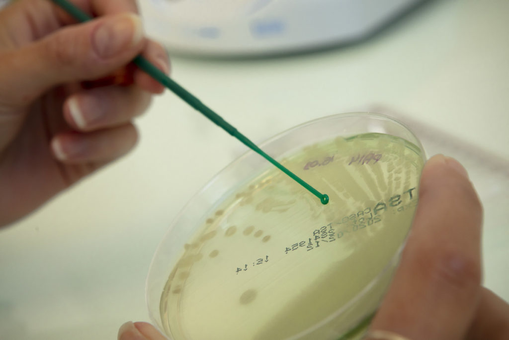
MICROBIOLOGY
Our microbiology platform allows us to handle microorganisms of various types: bacteria, yeast, molds, and phages, including a large number of pathogenic strains. Besides, the acquisition of an anaerobic station is a crucial element, allowing us to cultivate strains that have demanding atmospheric conditions.
In summary, the microbiology platform includes:
- 125 m² laboratory complying with Biosafety Level 2 (BSL2)
- Anaerobic station
- Analytical systems such as Tecan Spark 10M or BMG Labtech Spectrostar Nano microplate readers
- Microorganism counting systems (Bactobox and Spiral platter Eddy Jet)
- Electroporators
- Ultracentrifuge
- Climate-controlled chambers
- Remote Bruker® Maldi-Tof mass spectrometry platform
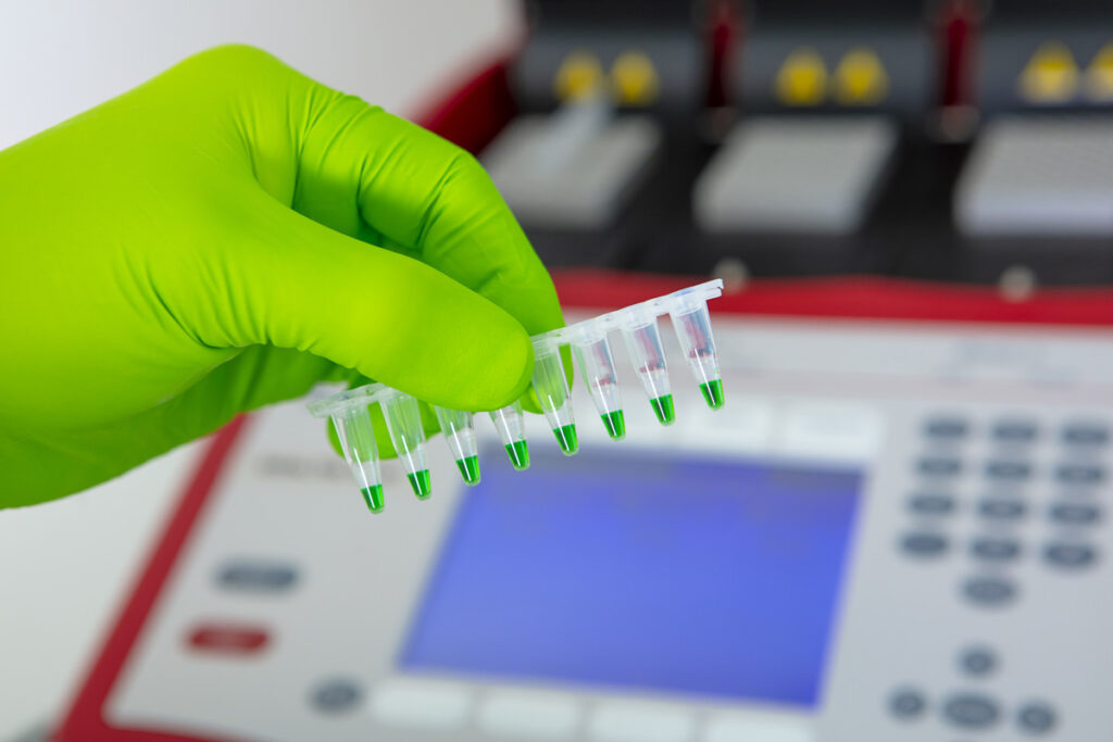
MOLECULAR BIOLOGY
Our molecular biology platform allows us to perform various missions such as genetic engineering or gene expression studies. This molecular biology technical platform complements the microbiology platform, as projects often require the use of both capabilities.
In summary, the molecular biology platform includes:
- 46 m² laboratory complying with Biosafety Level 2 (BSL2)
- Biometra Trio 48 Thermocyclers
- Real-time PCR systems
- Electrophoresis and DNA visualization systems
- Nucleic acid quantification systems
- Climate-controlled chambers
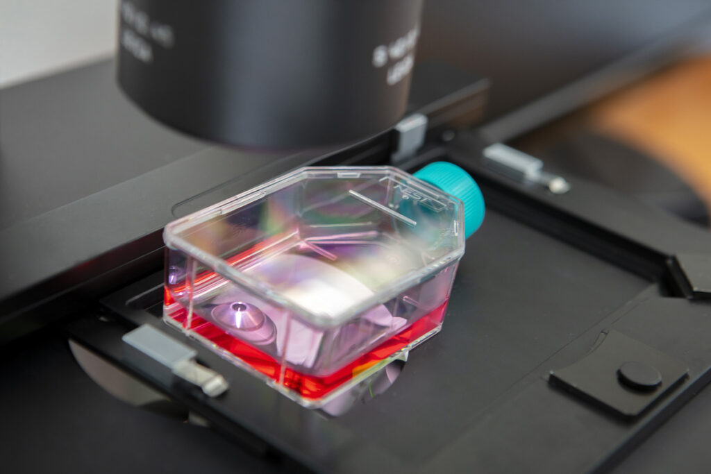
CELL BIOLOGY
Our cell biology platform enables us to handle different eukaryotic cell lines and expose them to microorganisms. Manipulating these cells allows us to offer cytotoxicity and cell viability evaluation services induced by compounds, bacteria, fungal microorganisms, or phages. Additionally, these models enable early-stage study of infection parameters. Eukaryotic cell lines are also useful for studying or producing intracellular bacteria.
In summary, the cell biology platform includes:
- 32 m² laboratory complying with Biosafety Level 2 (BSL2)
- Inverted fluorescence microscope
- Cell banks stored in liquid nitrogen – on a remote platform
- Tecan Spark 10M automated cell counting reader
Our Quality Requirements
These technical platforms are equipped with high-tech equipment validated by the laboratory’s teams, allowing us to provide high-quality services. Smaltis regularly acquires devices to meet the needs of new projects and stay at the forefront of technology.
Smaltis collaborates with qualified suppliers of materials, reagents, and equipment, with whom a relationship of trust has been established, some since the company’s inception. We are committed to using quality and robust equipment, essential for the smooth running of manipulations and the reliability of the results obtained. We carefully select the best to give you the best!
Our laboratory doors are always open; feel free to visit our premises and platforms!
An extensive network of partners and working groups
To provide turnkey solutions, Smaltis has surrounded itself with numerous partners. Smaltis is also a member of various networks to stay in touch with numerous stakeholders in the field of microbiology applied to health and to clearly understand the sector’s needs.

Joining POLEPHARMA means becoming part of a territory of shared needs, expertise, and solutions where everyone contributes to the excellence of all. Joining POLEPHARMA means participating in the economic and industrial network of stakeholders in the leading French pharmaceutical sector: Research laboratories, Production sites, Contract manufacturers, Suppliers, Training centers.
Read more

AFSSI, the French Association of Service and Innovation Companies for Life Sciences, was established in 2012 to bring together French companies providing services and technological innovation in the strategic field of Life Sciences. AFSSI aims to gather all sectors of biotechnology, chemistry, environment, cosmetology, agri-food, bioinformatics, including diagnostics and clinical trials.
Read more
Lyonbiopôle Auvergne-Rhône-Alpes, labeled as a competitiveness cluster at its creation, is the catalyst for the health innovation ecosystem in Auvergne-Rhône-Alpes, ensuring its connection, development, and promotion.
Read more

MabDesign is the French association for the industrial sector of biomedicines. Created in 2014 in response to the "Strategic Future Industrial Sector" call for projects, MabDesign aims to structure the industrial sector of biomedicines in France from R&D phases to market release, including bioproduction, and to generate the creation of innovative start-ups from French academic research.
Read more

PMT, a competitiveness cluster labeled by the French government in 2005, ws established as an Association uner the Law of 1901. Its mission is to catalyze innovation and accelerate the business of industrial companies in Bourgogne-Franche-Comté. PMT supports more than 240 industrial members specialized in specialty subcontracting or developing high-value-added products, offering daily and individualized support.
Read more

A group based on complementary expertise with multidisciplinary know-how. A range of integrated solutions in biotechnology. A complete platform, from DNA to recombinant antibodies. Customized services and products in Biotechnology: Molecular Biology – Immunology – Protein and Cell Engineering. An efficient synergy within a team of experts.
Read more

Our location
Smaltis established itself right from its inception in the heart of the TEMIS Santé zone in Besançon, built around the Jean-Minjoz University Hospital Center, the French Blood Establishment (Etablissement Français du Sang), and the National Institute of Health and Medical Research (INSERM). This business park was specifically designed to provide exceptional conditions to launch new ventures and aims to become a national hub for biomedical engineering. It is only logical that Smaltis positioned itself at the center of this dynamic network, aiming to be a genuine participant in ambitious undertaking, while being surrounded by partners focused on research and innovation in health, such as RD-Biotech, Skinexigence, Macopharma, Lymphobank, ISIFC, and Biotika Biomedical.
Get in touchAgustina LLANOS
Head of Biology
ANTABIO

We have collaborated with Smaltis on several occasions since 2014, notably as part of our ANT3273 project. This is an inhaled medicine targeting a key virulence factor of Pseudomonas aeruginosa involved in pathogenesis and would be an alternative or complement to existing antibiotic therapy for the treatment of P. aeruginosa infections in chronic respiratory diseases, such as bronchiectasis. Recently, Smaltis successfully completed a MLST sequencing project of a large number of strains with a very tight timeline to study the clonality of clinical isolates of P. aeruginosa from cystic fibrosis patients. The Smaltis team stands out for its expertise, proactive approach, and its communication and adaptability, which is why we continue to trust them.
Pierre BELICHARD
CEO
ENTEROME

As part of the development of our candidate EB8018 designed to block bacteria expressing virulence factor FimH (a key inducer of the inflammatory cascade of the intestine, particularly in patients with Crohn disease), we have successively called upon the services of SMALTIS. In the preclinical phase, they first quantified in vitro the adhesion and invasion capacities of a collection of strains isolated from patients on a model of intestinal cell infection and then evaluated the impact of our molecule on these strains. The quality of the work performed, the level of expertise of the team combined with the ability to listen and adapt, led us to include SMALTIS as a service provider in our Phase 1b clinical study. The team developed a customized procedure to isolate and dissociate bacteria called "adherent" and "invasive" in intestinal biopsies, and then demonstrated the action of our EB8018 compound on these strains. SMALTIS continues to support the development of EB8018 in a phase 2a clinical trial.
Romain DAILLERE
Head of Preclinical Research - Co-founder
EVERIMMUNE

As part of the Oncobax®-AK project and the development of Live Biotherapeutic Products to potentiate the efficacy of cancer immunotherapies, we called upon Smaltis' services to characterize one of our candidate bacterial strains. The objective was to provide a complete file to the regulatory authorities regarding the characteristics of the strain, before initiating clinical studies. The Smaltis team worked hard to develop the protocols and test conditions necessary to determine the specificities of the strain of interest. The rigor shown by Smaltis' researchers, the quality and detail of the data transmitted, as well as the scientific relevance of the various characterization tests, allowed us to meet the regulatory requirements and to initiate the clinical trials with confidence. We are delighted to have collaborated with Smaltis.
Maxime GUALTIERI
CSO
NOSOPHARM

As part of our anti-infective molecule developments, we have repeatedly sought the expertise of the Smaltis team for cloning and construction of specific mutants. The services provided by Smaltis have notably contributed to our study describing and characterizing new mechanisms of resistance to Odilorhabdines in Klebsiella pneumoniae, which was published in 2021. We know that we can rely on their know-how and expert scientific advice for our future developments.
Nicolas COULIN
Laboratory Manager
SOLICAZ
Solicaz develops natural bacterial biostimulation solutions to meet the challenges of agro-ecological transition and climate change. These PGPG (Plant Growth Promoting Rhizobacteria) bacteria provide benefits for improving plant growth and resistance. In order to exploit their full potential, we needed to call on a molecular biology expert to robustly determine the identity of a pool of selected strains. We identified Smaltis, who fully met our expectations by providing comprehensive expertise. The laboratory proved to be a true partner of choice, with whom we worked jointly to identify and test the method best suited to our needs. Their flexibility and commitment ensure that we have reliable results, and despite the geographical distance that separates us, our various subsidiaries will continue to work with Smaltis.
Eric ROBINET
Founder and President
LYMPHOBANK

Specialized in the production of cell banks from blood, for research and patient use, we needed to rely on a microbiology company to ensure the absence of contamination in our products from the start of our preparations. Established at the time in the premises of the EFS, it was only natural that we turned to Smaltis, located a few steps from our laboratories. Their work approach and the reliability of their results were exactly what we were looking for, and for several years now, we have entrusted them with all our microbiological analyses. The growth of our respective companies led us to look for new premises... and it is with pleasure that we took over the premises of Smaltis, who then moved into the building just across the street! It is important to have Smaltis in our network of reliable partners with common values, to contribute to the improvement of health!
Pascal MERTENS
R&D Director
CORIS BIOCONCEPT

As part of the development of rapid in vitro diagnostic (IVD) tools for detecting infectious pathologies in humans, we needed to evaluate the performance of new tests dedicated to the detection of bacterial antibiotic resistance mechanisms. We approached Smaltis, a company specializing in microbiology and molecular biology, and found them ideally suited to our needs. Smaltis made its expertise available to assess the efficacy and robustness of our tests on clinical strains with very specific antibiotic resistance profiles, compared with a molecular biology method. The results confirmed the sensitivity, specificity and accuracy of our test, and enabled us to meet regulatory requirements. For us, the Smaltis laboratory is an expert player on whom we can rely to support our developments and obtain the data we need to bring our tools to market.
Sophie MAC-MARY
CEO
SKINEXIGENCE

Smaltis is a laboratory that set up in the same building as our company, Skinexigence, from its inception. The proximity of our offices and the identification of a very interesting collaboration opportunity allowed us to quickly initiate a partnership to meet the growing demand from our clients for studies on the skin microbiota of volunteers. Specialized in the evaluation of the effectiveness of dermo-cosmetic products, it was only natural that we turned to Smaltis to provide our clients with expertise in the study of skin flora. For these studies, Smaltis performs skin sampling on our volunteers, sample preparation, and metagenomic analyses. The development of a very rigorous and calibrated protocol enables them to access relevant and reliable information regarding the evolution or not of the skin microbiota of a subject, in relation to the clinical parameters we observe.
Delphine GALLAND
Head of Preclinical Clinical Studies
VETOQUINOL

For the past 10 years, we have regularly turned to Smaltis for their services and expertise in microbiology, cellular biology, and molecular biology. The entrusted work focused on evaluating the efficacy and safety of various products related to infectious diseases. The smoothness of our interactions, the responsiveness of the entire team, and their adaptation to our regulatory requirements have allowed us to advance our research projects optimally. The trust we have placed in Smaltis over these 10 years has led us to include this provider among our preferred contacts.
Jérôme SCHMEISSER and Christelle IACONIS
Biology lab manager
and Scientist
DSM-FIRMENICH

In the context of our developments in animal health dietary supplements, we turned to SMALTIS to assess the impact of our products on the expression of genes of interest in different tissues. The molecular biology expertise of SMALTIS allows us to rely on this laboratory, whose qualitative results provide us with robust and reliable data. We also benefit from constructive exchanges with their teams to progress together throughout the project. SMALTIS thus constitutes a true extension of our R&D department, allowing us to focus on our own expertise while relying on this partner with whom we will continue to collaborate.
Caroline GEBUS
Bioprocess Scientist
OM Pharma

We use Escherichia coli strains to manufacture our Uro-Vaxom® product for the prevention of recurrent lower urinary tract infections and co-treatment of acute urinary tract infections and, in accordance with regulatory requirements, we must demonstrate the stability of our strains. We therefore asked SMALTIS to develop a tailor-made quality control method. In addition to their ability to listen to customer needs and propose new ideas, the SMALTIS team has also distinguished itself by the quality and accuracy of its analysis reports. At present, the partnership established with SMALTIS is a real asset for OM Pharma.
Laurence RINGENBACH
Cell Biology Lab Manager,
Immunodiagnostic Reagents,
DIACLONE SAS part of Medixbiochemica
Smaltis is a long-term partner with whom we have established a privileged relationship. We work in highly complementary domains and collaborate regularly to provide a comprehensive offering to our clients, utilizing microbiology and molecular biology services that we specifically require. We engage them for services such as sample contamination analysis, production of competent bacteria, and preparation of suspensions of inactivated strains for the development of new monoclonal antibodies. A true bond of trust has developed, facilitated by the geographical proximity of our respective locations and easy communication with their teams, whose flexibility, tenacity in problem-solving, and quality of work are crucial for us.
Olivier DHELLIN
Director of Pharmaceutical Development
NEOVACS
During our program to develop and industrialize the production of our Kinoïdes® vaccines, SMALTIS has proven to be a partner of choice for optimizing one of our cytokine production systems. Its service consisted in optimizing the genetic heritage of the host bacterium by excising DNA from exogenous sequences. Very attentive and with a rare expertise in microbiology, the SMALTIS team has always been benevolent and has never hesitated to guide us by providing sound advice. It is with pleasure that we will again call upon the services of SMALTIS to help us in our next challenges.
Eric SAMARUT
Co-founder and Scientific Director
OSTA THERAPEUTICS

Smaltis is an important partner for us, with whom we have established a close and early relationship to support the development of our new anti-infective molecules. We sought their recognized expertise in the field of antimicrobials and antibiotic resistance to support our proof of concept of the efficacy of our compounds, using specific strains from their mutant collection. With the help of growth kinetics, determination of Minimal Inhibitory Concentrations, and precise construction of mutant strains, along with their insightful advice and regular constructive exchanges, we obtained solid and essential elements to secure our data and continue our project.
Thomas GLOUDEN
PCR Development Expert
UNISENSOR

As part of the development of a simple, rapid molecular test for the detection of certain pathogenic micro-organisms in milk, we needed to work with a company with expertise in microbiology. We were looking for a qualified and experienced laboratory to prepare calibrated microbial suspensions and purified genetic material from different bacterial species. By identifying Smaltis, we were able to fully achieve our objectives thanks to the expertise of their team. We now regularly rely on this trusted partner for our recurring needs.
Clara LEANDRO
Head of Infection
TECHNOPHAGE

The advancement of Technophage's therapeutic pipeline products has received crucial support from SMALTIS in the scope of genetic modification of bacterial strains. Throughout this collaborative effort, the SMALTIS scientific team has demonstrated remarkable commitment to resolving various technical challenges, successfully aligning with our objective of acquiring a set of strains for our internal utilization, all within the stipulated timeline.